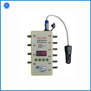
Model: SKX-1000E
SpO2 Function introduction:
1. External imitate finger,you can connect to any SpO2 test instruments easily;
2. It is a multi-functional optical simulator, built-in frequently-used BCI and Nellcor wave-forms curv
e;
3. The Oximetry simulate range:80%,85%,90%,98%, these 4 points value detection calibration,tole
rance<=1%;
60%,65%,70%,75%,these 4 points value detection calibration, tolerance<2%;
4. Pulse rate simulate range:30,60,80,100,120,160,180,240 bpm total 8 test points, tolerance<=1b
pm;
ECG function introduction:
1. Normal ECG waveform, 12-lead different ranges and types synchronous waveform;
2. Plus and reserve direction heart rate detection waveform;
3. Square wave, used the square wave to measure scan speed;
4. Sinusoidal wave, measure amplitude-frequency characteristic
5. Can change the T-wave amplitude, heart rate value, R-wave amplitude, and R-wave width of an
alog QRS waveform;
6. The Respiration rate of breathing waveform can be changed+ ECG waveform;
7. You can set the ECG signal amplitude;
Waveform code introduction:
1. Oxygen saturation values, set the SpO2 rate of simulator;
2. Pulse rate value, set the simulator pulse rate value;
3. Select the data curve of Oxygen saturation;
4. Normal ECG waveform, 12-lead different ranges and types synchronous waveform;
5. Plus-minus triangular waveform;
6. Square wave, used the square wave to measure scan speed;
7. Sinusoidal wave, measure amplitude-frequency characteristic
8. Can change the T-wave amplitude, heart rate value, R-wave amplitude, and R-wave width of an
alog QRS waveform;
9. Simulate Respiration waveform;
A. Set the amplitude of signal;
Note:
1.The simulator will automatically generate a value 98 of blood Oxygen, 80 of pulse rate, blood Ox
ygen parameter data of NELLCOR curve,
and code as 4 normal ECG waveform, and respiration rate as 15bpm respiratory waveform.
2. The imitate finger part, against the LED of the Spo2 sensor; and the imitate finger part, against
the SpO2 sensor PD; and make sure
that the white color window of imitate finger stay in line with the PD of SpO2 sensor, or it will lead t
o invalid data.
3. The simulator will automatically generate lead II amplitude at 1mV,and in the heart rate detectio
n wave, square wave, sinusoidal wave,
QRS wave,all amplitude of lead II is 1mV.
4. When the battery falls lower than a certain value, the contents of the digital display will flash to i
ndicate low battery, and then continue
to work only about four hours. Please note that at this moment the ECG waveform amplitude will d
ecrease.
5. The ECG part connection to ECG machine: RA-R(right arm), LA-L(left arm), LL-F(left leg),RL-R
F(right leg), C1-C6(chest lead).
6. The ECG part connection to patient monitor:RA-right arm(white), LA-left arm(black), LL-left leg(r
ed), RL-right leg(green), C1-C6 chest lead(brown).
7. ECG part 3 leads connection: RA-right arm(white), LA-left arm(black), LL-left leg(red).
8. Different identify corresponding connection:L-LA, R-RA, RF(N)-RL, F-LL, C-V.
Key description:
There are 4 keys, followed by selection keys, increase key, reducing key, the Enter key, in addition
to a set of key combinations
Select Key:
This key is used to select the parameter you want to change; there are 4 LED tubes to display the f
our codes represent what is displayed,
The 1st LED representative waveform code, LED 2-4 representatives the parameter (2 is the value
of one hundred, 3 representatives of ten, 4 representatives of bits)
Increase key:
Use it to increase the parameter
Reducing key:
Use it to reduce the parameter
Enter key:
Use it to confirm the parameter you selected
Combination key:
Press the select key and Enter key at the same time, release the Enter key, the release the select
key,
double-click the Enter key again, the combination key was selected successfully, and it will display
different content.
The operation ways of each code:
1. Blood Oxygen value:
1) The blood Oxygen value setting at:80%,85%,90%,98%, these 4 points value detection calibrati
on,tolerance<=1%;
At 60%,65%,70%,75%,these 4 points value detection calibration, tolerance<2
%;
2) Initial value:98%; It can be select and set the blood oxygen value with increase key and reduce
key directly.
2. Pulse rate value:
1) Pulse rate value setting at: 30,60,80,100,120,160,180,240 bpm total 8 test points;
2) Tolerance<=1 bpm;
3) Initial value is 80 bpm,It can be select and set the pulse rate value with increase key and reduc
e key directly.
3. Waveform data curve selection:
Data 1 is BCI curve, data 2 is NELLCOR curve.
4. Normal ECG waveform:
* Heart rate setting range: 10-200bpm (Initial value: 60 bpm)
* Signal amplitude is fixed
5. Plus-minus triangular waveform
* Frequency range: 10-300bpm (Initial value: 75 bpm)
* Amplitude range: 0.1-4Mv (10:0.1mV, 400:4mV)
* 2 modes, Mode 1: Positive waveform; mode II: negative waveform; Use combinations key to
choose.
6. Square wave
* Frequency range: 0.1Hz-10Hz (10:0.1Hz, 100:10Hz); (Initial value: 1 Hz )
* Amplitude range: 0.1-4Mv (10:0.1mV, 400: 4mV);
7. Sinusoidal wave
* Frequency range: 1-100Hz (Initial value: 25 Hz)
* Amplitude range: 0.1-4Mv (10:0.1mV, 400:4mV)
8. Analog QRS-T waveform:
This waveform have 4 operating modes,
Model 1: the T-wave amplitude can be changed;
Model 2: Set the waveform frequency;
Mode 3: setting the amplitude of QRS waveform;
Model 4: Set the width of QRS wave;
the operation mode can change with combination keys.
* T wave amplitude setting range: 0.1mV-2mV; (Initial value: 10)
* Waveform frequency setting range: 20-300bpm; (Initial value: 75)
* QRS waveform amplitude range: 0.1mV-2mV;(Initial value: 1mV)
* QRS waveform width range: 10ms-150ms;(Initial value: 80ms)
9. Simulate respiration wave:* Frequency range: 10-100time/min; (Initial value: 15bpm)
* Note: the respiration lead is RA-LL, baseline impedance is 1k.If the patient monitor respiration le
ad is other lead, please set the parameter of monitor or change the lead connect way.
A. Signal amplitude setting:
* Amplitude range: 0.1mV-4mV (10:0.1mV, 400:4mV); (Initial value: 100)
* Note: this amplitude setting will affect the 5,6,7 waveform amplitude.